What Is CBCT Imaging and Why Would Your Dentist in Frisco Recommend It?

Most dental concerns can be evaluated with an examination and standard X-rays. But sometimes, your dentist needs to see more than a flat image can show.
CBCT imaging creates a three-dimensional view of the teeth, jawbone, tooth roots, nerves, sinuses, and nearby structures. That added detail can be especially helpful when planning dental implants, evaluating impacted teeth, investigating unexplained pain, or preparing for more complex care.
What Happens During a CBCT Scan?
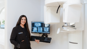
CBCT stands for cone beam computed tomography. During the scan, the machine moves around your head and captures images from several angles. Those images are then combined into a detailed 3D model that your dentist can review from different directions and in individual sections.
The appointment itself is usually straightforward. You remain still while the scanner rotates around your head, without touching you. The process is quick, and the images are often available for your dentist to review right away.
What makes CBCT different is depth. A standard dental X-ray can show the teeth and surrounding bone in two dimensions. A CBCT scan allows your dentist to examine height, width, position, and the relationship between structures that may overlap on a traditional image.
That can make a meaningful difference when treatment involves a nerve, the sinus, limited bone, unusual root anatomy, or an area that has been difficult to diagnose.
Why Your Dentist May Recommend One
A CBCT scan is not part of every dental visit. It is generally recommended when the additional information could improve the diagnosis or help your dentist plan treatment more carefully.
Dental implant planning is one of the most common reasons. Before placing an implant, your dentist needs to know how much bone is available and where important structures are located. The scan can help identify a suitable implant position and show whether bone grafting may need to be considered.
CBCT imaging may also be useful before removing an impacted wisdom tooth or another tooth with complex roots. Seeing how those roots relate to nearby nerves, teeth, and sinus spaces helps your dentist prepare for the procedure and explain any relevant risks beforehand.
In other cases, the reason for the scan is less obvious. A patient may have ongoing pain, swelling, a suspected root fracture, or an infection that does not appear clearly on standard X-rays. Three-dimensional imaging can sometimes reveal details that were hidden or difficult to interpret from a single angle.
For root canal treatment, restorative care, clear aligners, or bite and jaw concerns, the scan may help the dental team understand how several structures are working together before deciding on the treatment sequence.
What Should You Expect?
Before the scan, you may be asked to remove glasses, jewelry, removable dental appliances, or other metal objects that could affect the image. Let the dental team know if you are pregnant or think you may be pregnant.
Once the scan is complete, your dentist can walk you through the findings and explain why they matter. You may be shown the amount of available bone, the location of an infection, the path of a nerve, or another detail that could influence your care.
The scan provides information. Your dentist still considers your symptoms, examination findings, health history, and treatment goals before making a recommendation.
At brush365, CBCT imaging is used when a more complete view can support accurate diagnosis and thoughtful treatment planning. If you’re in Frisco, you can visit our Frisco dental office or contact us to discuss your dental concern and learn whether three-dimensional imaging would be beneficial for your care.